Personal Details
|
Name: Date of Birth: e-mail: Web: Phone: Nationality: Languages: Occupation: |
Stefan Schenke 28th May 1978 stefan_schenke@yahoo.de www.stefan-schenke.net/cv +49 179 32 82 795 German German, English MSc. Student in Computer Science |
Education
| October 1998 to July 2005: | Study of MSc. in Computer Science with
minor subject Medical Science at the Friedrich-Schiller-University in Jena, Germany |
|
December 2004 to June 2005: |
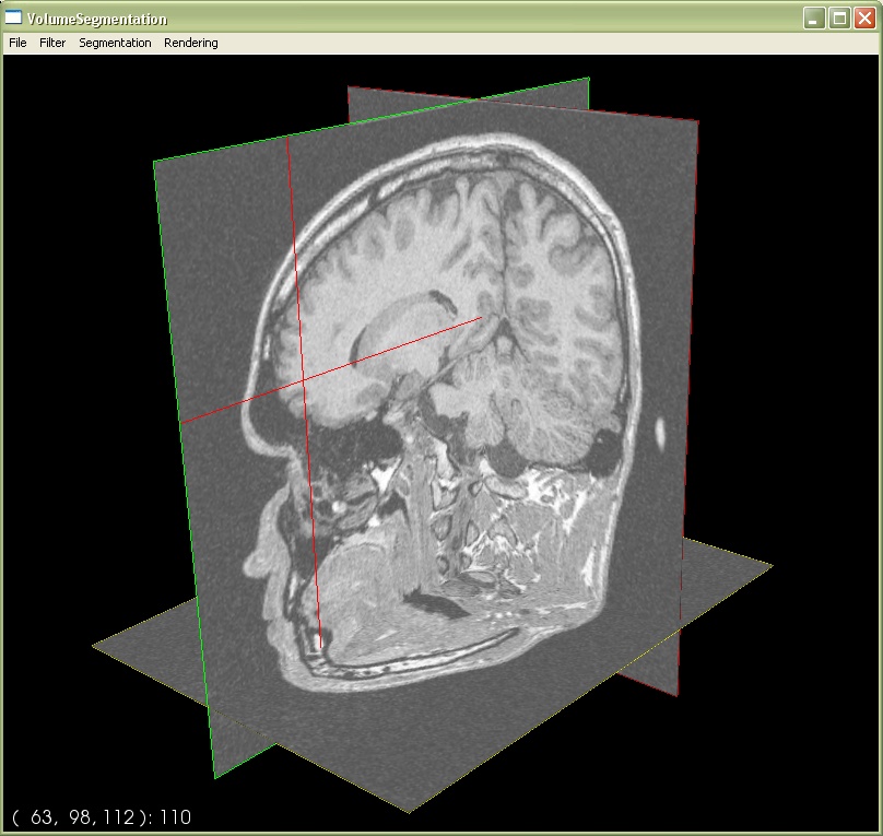
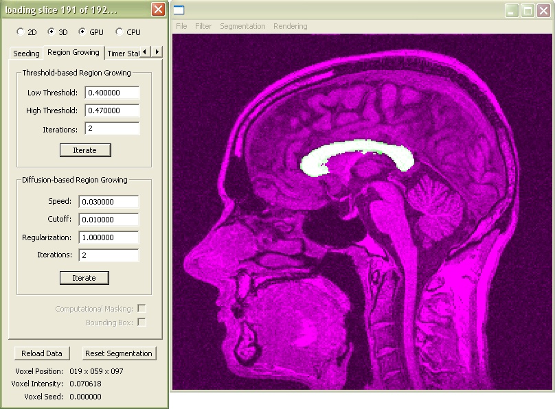
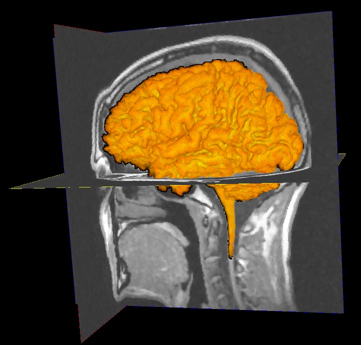
Preparation of MSc. Thesis "Analysis and Design of GPU-based Algorithms
for Interactive Volume Segmentation" at the University of Auckland, New Zealand |
|
Taken Computer Science courses including:
|
||
| February 2001: | Associate Degree in Computer
Science from the Friedrich-Schiller-University
in Jena, Germany |
Work Experience
| November 2005 to April 2007: |
Research Associate at the Department for Experimental
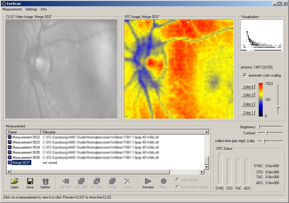
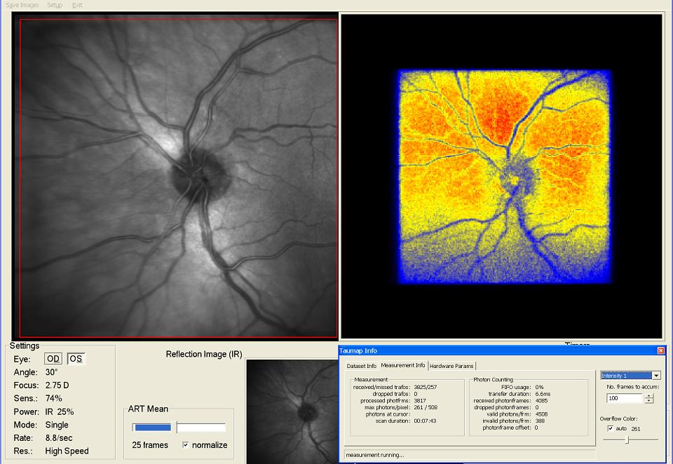
Ophthalmology at the Friedrich-Schiller-University, Jena, Germany. In this project an industrial Laser Scanning Ophthalmoscope has been advanced to allow for 2-dimensional Fluorescence Lifetime Imaging (FLIM) at the human ocular fundus. The method of Time Correlated Single Photon Counting is used in conjunction with a sensitive detector to determine the decay-times of the natural fluorophores. The aim of this project is to measure certain parameters of metabolism in the retinal tissue and detect diseases like age-related macula degeneration (AMD), diabetic retinopathy or glaucoma before pathological changes are manifest. Please see publications for further details. Main achievements in this research group:
Head of Research Group: HDoz. Dr.-Ing. habil. Dietrich Schweitzer |
|
|
| December 2004 to June 2005: | Preparation of MSc. Thesis "Analysis and Design of GPU-based Algorithms
for Interactive Volume Segmentation" at the University of Auckland, New Zealand
Supervised by Prof. Denzler and Burkhard Wünsche, PhD |
|
|
| May 2004 to October 2004: | Part-time based work in the Software Engineering Department
of Carl Zeiss Meditec in Jena, Germany.
|
 |
|
| March 2002 to July 2004: |
Collaboration in a research project at the Department for Experimental
Ophthalmology at the Friedrich-Schiller-University, Jena, Germany. In this project a Confocal Laser Scanning Ophthalmoscope has been advanced to measure the 2-dimensional time-resolved autofluorescence of the ocular fundus. The method of Time Correlated Single Photon Counting is used in conjunction with a sensitive detector to determine the decay-times of the natural fluorophores. The aim of this project is to measure certain parameters of metabolism in the retinal tissue and detect diseases like age-related macula degeneration (AMD), diabetic retinopathy or glaucoma before pathological changes are manifest. This research project is partially funded by the Federal Ministry for Education and Research. I have contributed to the development of a measurement-technique which led to the application of a patent. Please see publication-section for further details and achievements. My assignment was the development of the measurement software "EyeScan" with the following features:
Supervised by HDoz. Dr.-Ing. habil. Dietrich Schweitzer |
 |
|
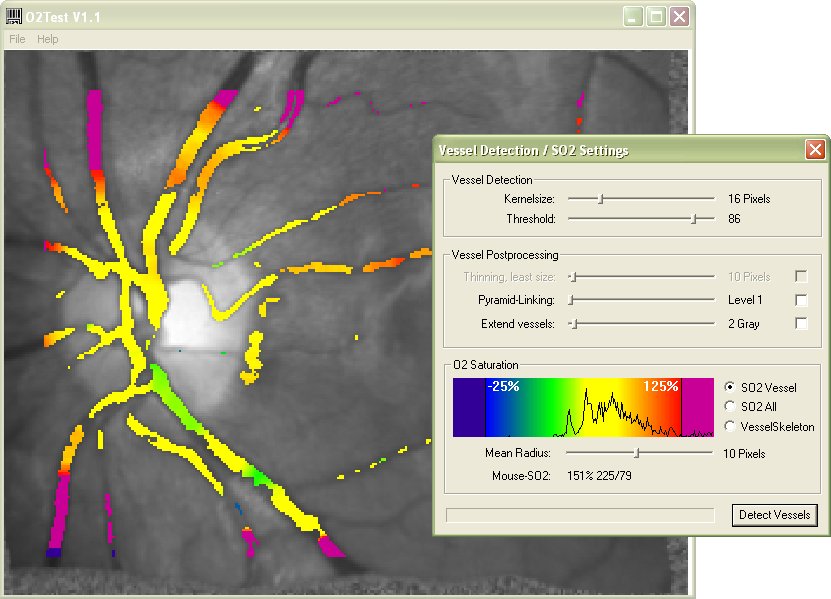
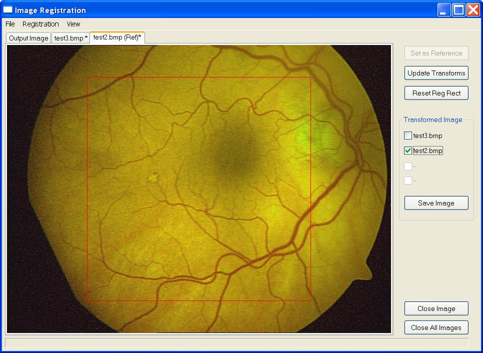
| September 2003 to January 2004: | Development of the software VesselO2 for a research
project at the Department for Experimental Ophthalmology at
the Friedrich-Schiller-University, Jena, Germany. It is used to compute the
oxygen saturation in the retina from images taken by a fundus camera at
different wavelengths of the reflected light.
Under direction by HDoz. Dr.-Ing. habil. Dietrich Schweitzer |
 |
|
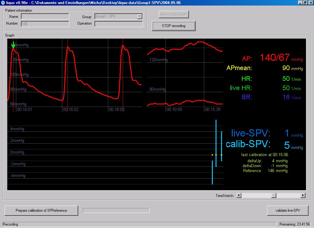
| March to June 2002: | Development of the medical data-acquisition and
-visualization software "Liquo" in conjunction with the MD thesis of a medical
science student at the Department of Anesthesiology and Intensive Care Medicine
of the Friedrich-Schiller-University, Jena, Germany.
Supervised by Samir Sakka, MD, PhD |
|
|
| August to October 2001: | Internship at Vulpine 3D Technologies GmbH
which evolved to Trinigy GmbH, Reutlingen, Germany.
References provided on request |
 |
|
| May 1998 to June 2001: | Student Assistant at the Fraunhofer Institute
for Applied Optics and Precision Engineering, Jena, Germany.
References provided on request |
 |

Referees
| HDoz. Dr.-Ing. habil. Dietrich Schweitzer | Research Department Experimental Ophthalmology at the Friedrich-Schiller-University Hospital, Jena, Germany Dietrich.Schweitzer@med.uni-jena.de Phone: +49 3641 933027 |
|
| Burkhard Wünsche, PhD | Graphics Group / Biomedical Imaging & Visualization Department of Computer Science University of Auckland, Auckland, New Zealand burkhard@cs.auckland.ac.nz Phone: +64 9 3737 599 x83705 |
|
| Samir Sakka, MD, PhD | Department of Anesthesiology and Intensive Care
Medicine of the Friedrich-Schiller-University Hospital, Jena, Germany Samir.Sakka@med.uni-jena.de Phone: +49 3641 9 32 22 48 |
Publications
Publications
-
In vivo autofluorescence lifetime imaging at the fundus of the human eye
Dietrich Schweitzer, Martin Hammer, Frank Schweitzer, Stefan Schenke, Eckhard Birckner, Wolfgang Becker, Axel Bergmann
Proceedings of SPIE Vol. 6138 (2006) 613808-1 – 613808-10
-
GPU-Based Volume Segmentation
Stefan Schenke and Burkhard C. Wünsche
Image and Vision Computing New Zealand, Dunedin, 28.-29. November 2005
-
Comparison of time-resolved autofluorescence in the eye-ground of healthy subjects and patients suffering from age-related macula degeneration
D. Schweitzer, F. Schweitzer, M. Hammer, S. Schenke, S. Richter
Progress in Biomedical Optics and Imaging, Vol. 6, No. 31, SPIE 5862, 58620 R-1 – 58620 R-12, 2005
-
Time-Resolved Autofluorescence Yields More Information Than Intensity Measurements of Fundus Autofluorescence
D. Schweitzer, F. Schweitzer, M. Hammer, S. Schenke, S. Richter
Invest. Ophthalmol. Vis. Sci. 46: E-Abstract 227, 2005
-
In vivo measurement of time-resolved autofluorescence at the human ocular fundus
D. Schweitzer, M. Hammer, F. Schweitzer, R. Anders, T. Doebbecke, S. Schenke, E. R. Gaillard
Journal of Biomedical Optics, Vol. 9 No. 6 pp 1214-1222, 2004
-
Veränderungen der Autofluoreszenzlebensdauer am Fundus
nach Sauerstoffprovokation
(Alteration of autofluorescence decay time in the fundus after breathing 100% oxygen)
Schweitzer D., Hammer M., Anders R., Doebbecke T., Schenke S.
Ophthalmologe 2004, 101:66-72
-
Metabolic mapping by time-resolved autofluorescence
Schweitzer D., Hammer M., Schenke S., Anders R.
European Journal of Ophthalmology Vol. 13 No. 2, 2003, p 230
-
Evaluation of time-resolved autofluorescence images of
the ocular fundus
Schweitzer D., Hammer M., Schweitzer F., Schenke S., Gaillard E.R.
Proceedings of SPIE 5141 "Diagnostic Optical Spectroscopy in Biomedicine II" ed. by G.A. Wagnieres, 2003 pp 8-17
Talks
- Steuerung der intraoperativen Volumentherapie an Hand der systolischen Druckvariation: Gibt es Vorteile hinsichtlich der Organfunktionen?
M. Büttner, S. Schenke, O. Thümer, W. Schummer, S. G. Sakka
Deutscher Anästhesiecongress, München, 16.–19. April 2005
- Systolic pressure variation guided volume therapy – are there any advantages in organ function?
O. Thümer, M. Büttner, S. Schenke, W. Schummer, S. G. Sakka
European Society of Intensive Care Medicine - 18th Annual Congress, Amsterdam, 25.–28. September 2005
- Comparison of time-resolved autofluorescence in the eye-ground of healthy subjects and patients suffering from age-related macular degeneration
D. Schweitzer, F. Schweitzer, M. Hammer, S. Schenke, S. Richter
European Conference on Biomedical Optics, München, Mai 2005-10-04
- Interpretation der dynamischen Autofluoreszenz am Augenhintergrund
D. Schweitzer, F. Schweitzer, M. Hammer, S. Richter, S. Schenke
XIV. Workshop „Okuläre Mikrozirkulation“, Weimar, 26.–27. Februar 2005
- Diagnostische Möglichkeiten der zeitlich und spektral aufgelösten Autofluoreszenzbeobachtung bei alterbedingter Makuladegeneration
M. Hammer, D. Schweitzer, S. Richter, S. Schenke
13. Tagung der Gesellschaft der Augenärzte Sachsen-Anhalts und Thüringens, Jena, 12.–13. November 2004
- Erste Studienergebnisse zur Messung der 2-dimensional zeitaufgelösten Autofluoreszenz am Augenhintergrund
D. Schweitzer, F. Schweitzer, S. Schenke, M. Hammer, S. Richter
XIII Workshop „Okuläre Mikrozirkulation“, München, 28.–29. Februar 2004
-
Evaluation of time-resolved autofluorescence images of the ocular fundus
Schweitzer D., Hammer M., Schweitzer F., Schenke S., Gaillard E.R.
European Conference on Biomedical Optics June, 22-25, 2003, Munich
-
Metabolic mapping by time-resolved autofluorescence
Schweitzer D., Hammer M., Schenke S., Anders R.
International Workshop NTG & PEX, April 3-5, 2003, Erlangen, Germany
-
Metabolic mapping by time-resolved autofluorescence
Schweitzer D., Hammer M., Schenke S., Anders R.
3. CAFIA meeting Computer Assisted Fundus Image Analysis, March 28-30, 2003, Torino, Italy
- Änderung der zeitaufgelösten Autofluoreszenz nach Sauerstoffprovokation
D. Schweitzer, M. Hammer, S. Schenke, P. Gänßler
Workshop "Okuläre Mikro- und Makrozirkulation" Dresden, 28. Februar – 01. März 2003
Pending Patent
-
Verfahren und Anordnung zur Gewinnung und Auswertung kontrastreicher
Bilder der zeitaufgelösten Fluoreszenz von bewegten Objekten, beispielsweise
des Augenhintergrundes
(Method and arrangement for acquisition and evaluation of contrast-rich images of time-resolved fluorescence of moving objects, e.g. the ocular fundus)
Schweitzer D., Schweitzer F., Schenke S., Hammer M.
10. February 2004, Aktenzeichen 102004006960.3
Skills
Programming:
-
Several years experience in Object Oriented Software Development in C/C++
using Microsoft Visual Studio .NET -
Developing Graphical / Image Processing Applications using
-
GPGPU-Development
Implementing Image Processing Tasks on GPUs -
Knowledge of Win32-API including
GUI / Controls / Windowing / GDI / GDI+ / Multithreading - Basic knowledge in Matlab, JAVA, PASCAL, VisualBasic, HTML 4.0, CSS
General:
- MS Windows XP, 2000, 98
- MS Office including database design and VBA-macro-programming in MS Access
- basic Unix/Linux
Personal:
- ability to work in a team environment as well as self-dependent
- research experience
- working reliably and precisely
- ability to learn quickly new methods and techniques
- being versatile and flexible